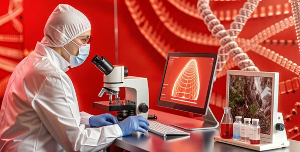
Understanding Histoblur: A Revolutionary Tool for Histology

In the ever-evolving field of medical research, the integration of technology has been crucial in enhancing the capabilities of researchers and healthcare professionals. One of the standout innovations in this arena is Histoblur. This article explores what Histoblur is, its significance in histology, its key features, and how it is transforming medical research and diagnostics.
What is Histoblur?
Histoblur is an advanced imaging and analysis software specifically designed for histology, the study of microscopic tissues. It aims to streamline the process of analyzing histological images, making it easier for researchers and pathologists to interpret complex data. Histoblur integrates state-of-the-art algorithms and machine learning techniques to provide users with insightful analytics, facilitating more accurate diagnoses and research outcomes.
The Importance of Histology
Histology plays a vital role in understanding disease mechanisms, diagnosing conditions, and developing new treatments. The examination of tissue samples under a microscope allows healthcare professionals to identify abnormalities, such as cancerous cells or inflammatory conditions. However, analyzing histological images can be time-consuming and prone to human error. Histoblur addresses these challenges by automating various aspects of the analysis process.
Key Features of Histoblur
Histoblur stands out in the field of histology due to its comprehensive set of features. Here are some of the most significant aspects that make Histoblur an essential tool for researchers and pathologists:
1. Automated Image Analysis
One of the most impressive features of Histoblur is its automated image analysis capabilities. Using advanced algorithms, Histoblur can quickly and accurately analyze large sets of histological images. This automation reduces the time researchers spend on manual analysis, allowing them to focus on more critical aspects of their work.
2. High-Resolution Imaging
Histoblur supports high-resolution imaging, which is crucial for detailed analysis of tissue samples. The ability to capture and analyze images at a granular level enables researchers to identify minute changes in tissue structure, which can be essential for accurate diagnoses and understanding disease progression.
3. Machine Learning Integration
Histoblur employs machine learning techniques to enhance its analytical capabilities. By training on vast datasets, the software learns to recognize patterns and anomalies in histological images. This feature not only improves accuracy but also aids in the development of predictive models that can foresee potential health risks based on tissue changes.
4. User-Friendly Interface
Despite its advanced capabilities, Histoblur is designed with user-friendliness in mind. The intuitive interface allows users to navigate the software effortlessly, even if they have limited technical expertise. This accessibility makes Histoblur suitable for a wide range of users, from experienced researchers to medical students.
5. Comprehensive Reporting Tools
Histoblur includes robust reporting tools that generate detailed analytics and visualizations. Users can easily create reports summarizing their findings, complete with graphs and charts that illustrate key data points. This feature is invaluable for presenting research results in academic or clinical settings.
The Significance of Histoblur in Medical Research
Histoblur’s impact on medical research is profound, offering several key benefits that enhance both the quality and efficiency of histological studies.
1. Improved Accuracy
The integration of automated analysis and machine learning significantly reduces the likelihood of human error in interpreting histological images. By providing accurate and consistent results, Histoblur enhances the reliability of research findings and clinical diagnoses.
2. Enhanced Productivity
Researchers often face tight deadlines, and the time-consuming nature of manual image analysis can hinder productivity. Histoblur accelerates this process, allowing researchers to analyze large datasets in a fraction of the time it would take manually. This boost in productivity is particularly beneficial in high-stakes environments, such as clinical laboratories.
3. Facilitating Collaborative Research
Histoblur promotes collaboration among researchers by providing a centralized platform for sharing and analyzing histological data. Teams can work together more effectively, accessing and analyzing the same datasets, which fosters a more collaborative research environment.
4. Accelerating Drug Development
In pharmaceutical research, understanding the effects of new drugs on tissues is critical. Histoblur helps researchers analyze how drugs interact with tissues at a microscopic level, facilitating the development of more effective treatments. This acceleration in research can lead to faster clinical trials and, ultimately, quicker patient access to new therapies.
Challenges in Histology and How Histoblur Addresses Them
Despite its significance, the field of histology faces several challenges that Histoblur aims to mitigate.
1. High Volume of Data
Histology generates vast amounts of data that can be overwhelming for researchers to manage. Histoblur’s automated analysis features help tackle this issue by processing large datasets quickly and efficiently. This capability ensures that researchers can keep up with the demands of modern histological studies.
2. Variability in Interpretation
Interpreting histological images can vary significantly among different pathologists, leading to inconsistent results. Histoblur’s machine learning algorithms help standardize the analysis process, reducing variability and ensuring that interpretations are consistent across different users.
3. Resource Limitations
Many research facilities may lack the resources to invest in expensive imaging equipment or dedicated staff for image analysis. Histoblur provides an accessible solution that leverages existing imaging technologies, allowing facilities to enhance their capabilities without significant financial investment.
Case Studies: Histoblur in Action
To illustrate the transformative potential of Histoblur, let’s explore some hypothetical case studies in various fields:
Case Study 1: Cancer Research
In a leading cancer research institute, researchers utilized Histoblur to analyze tissue samples from clinical trials. By automating the analysis of tumor samples, the team was able to identify specific markers associated with treatment responses. This information was crucial in refining treatment protocols and personalizing patient care.
Case Study 2: Infectious Disease Studies
A team studying infectious diseases employed Histoblur to analyze tissue samples from patients with chronic infections. The automated image analysis allowed researchers to track changes in tissue structure over time, providing insights into disease progression and treatment efficacy. This data was invaluable in developing targeted therapies for patients.
Case Study 3: Education and Training
In a medical school, Histoblur was integrated into the histology curriculum. Students used the software to analyze tissue samples, gaining hands-on experience with advanced imaging techniques. This exposure enhanced their learning and prepared them for future roles in clinical practice and research.
The Future of Histoblur
As technology continues to advance, the future of Histoblur looks promising. Here are some potential developments to watch for:
1. Enhanced Machine Learning Capabilities
As more data becomes available, Histoblur’s machine learning algorithms can be refined to improve accuracy and predictive capabilities. Future versions of the software may incorporate deeper learning techniques, enabling even more sophisticated analyses.
2. Integration with Other Technologies
Histoblur may expand its capabilities by integrating with other technologies, such as artificial intelligence and augmented reality. These enhancements could further streamline the analysis process and improve the visualization of histological data.
3. Expansion into New Fields
While Histoblur is primarily focused on histology, its underlying technology could be adapted for use in other fields, such as forensic science or veterinary medicine. This diversification could open new avenues for research and application.
4. User Community and Collaboration
Building a user community around Histoblur could foster collaboration and knowledge sharing among researchers. Online forums, webinars, and training sessions could enhance user engagement and facilitate the exchange of best practices.
Conclusion
Histoblur represents a significant advancement in the field of histology, providing researchers and healthcare professionals with a powerful tool for image analysis and data interpretation. With its automated capabilities, machine learning integration, and user-friendly design, Histoblur is transforming the way histological studies are conducted.
More Read
More Read
Pest-Free Living: Trust Carolina Exterminating for All Your Needs


Responses